It�s bad enough when a process glitch leads to a failure caught by end-of-line electrical testing. But the more insidious worry for engineers is the invisible anomaly that slides through the tests and can cause an electrical failure of the product at some time in the future�perhaps in a month, six months, or a year. These are the anomalies that may lead to massive, well-publicized recalls that can damage a company�s reputation.
Many of these randomly occurring electrical failures are triggered by anomalies in the plastic-encapsulated IC package and have nothing to do with the silicon itself. The anomalies are cracks, delaminations, and voids in the package that either expand through normal thermal cycling or collect water and contaminants that permit corrosion.
A quietly growing trend gives manufacturing engineers a relatively painless way to find at least some of these anomalies before they can do any real harm. Developed and patented by Sonoscan, the method uses an acoustic microscope but does not, at least initially, make any acoustic images.
Instead, a component, generally a plastic-encapsulated IC, is scanned acoustically before it is surface mounted (Figure 1). The purpose of the scan is to collect all of the acoustic data as waveforms from the entire volume of the component.
To do this, the area of the component is scanned multiple times at increasing depths. In theory, it might be possible to obtain the acoustic data with a single scan of the component�s area, but trying to cover the entire depth of the component in one shot leads to distortions, and the value of this method lies in its precision.
The acoustic data is stored electronically, and the component is surface mounted. The acoustic data typically is ignored unless the component fails.
When a failure is found electrically, the stored electronic data is used to make acoustic images from the component. One estimate is that 20,000 different images theoretically can be made from the electronic data, although in practice, half a dozen images may be enough to pinpoint the cause of the failure. The data file is scanned to make acoustic images as though it were the physical component although the actual component may already have been destroyed by testing.
The nature of the electrical failure and their own knowledge of the component may lead engineers to look first at acoustic images of the die face, for example, or of the die attach material or the lead fingers. Often, they are looking for small, incipient anomalies that could expand during reflow and particularly during lead-free reflow.
The stored acoustic data permits engineers to see the internal structure of the component in its original, pristine state as it existed when it was delivered from the supplier. Engineers might find, for example, that the component originally had a very small delamination between the molding compound and the die face at one corner of the die. Such anomalies are fairly common and likely to expand�perhaps slowly, perhaps rapidly�until they snap off a wire bond on the die face.
If engineers find such a die-face delamination in the acoustic data, and if the post-reflow component has an even larger die-face delamination along with the electrical failure, then engineers have rapidly identified a significant process failure. It may be worthwhile for engineers to examine the entire lot of components that included the failed component. This will allow them to look for additional small die-face delaminations that might fail electrically right after reflow or which might fail electrically weeks or months later. Using the stored acoustic data lets engineers discover the root cause quickly.
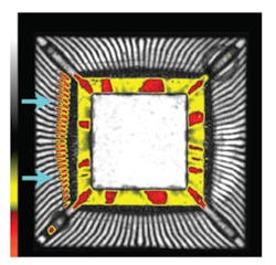
Figure 2 is the acoustic image of a thin quad flat pack (TQFP) that experienced an electrical failure after reflow. The acoustic image was made not from the TQFP itself but from the stored acoustic data.
Acoustic images, whether made from a physical component or from a stored file, generally are gated on a particular depth; that is, only the return echoes from the depth of interest are used to make the acoustic image. In Figure 2, the gate included the lead fingers, the die, and the top of the die paddle. Red and yellow areas in the acoustic image indicate gap-type anomalies such as delaminations, cracks, and voids.
The exception in this image is the square yellow region surrounding most of the die. Yellow in this location identifies the wires that extend from the die to the lead fingers. Because they are curved, the wires scatter ultrasound instead of reflecting it in a meaningful manner. For that reason, the yellow of the wires does not indicate an anomaly.
Between the groups of wires, however, are areas of red. In these areas, ultrasound is traveling slightly deeper into the virtual component and being reflected from the depth at which the molding compound meets the outer portion of the die paddle.
The red color indicates that the molding compound is separated from the die paddle. This generally is not considered serious; there is little danger from expansion of the delamination unless it runs under the die and delaminates the die attach layer.
Near the left edge of the component is another area of red, right at the inner ends of the lead fingers. Here red indicates small delaminations on the lead fingers.
The delaminations are in the same area where the lead wires are bonded to the lead fingers. Either expansion or corrosion within a delamination can break the wire. And a broken wire on one or more of these lead fingers probably was the cause of the electrical failure found by end-of-line testing.
The quick evaluation of the acoustic images made from the data file gave engineers a good starting point for finding and eliminating this process flaw. The delaminations on the lead fingers might be the result of surface contamination or of an improper plating material. If surface contamination is the cause, it also could explain the delamination seen on the outer part of the die paddle.
It is worth noting that the ultrasound pulsed into a sample by the scanning transducer of an acoustic microscope is reflected only from the material interfaces inside the sample. The ultrasound travels through the molding compound, which is homogeneous, without sending back any echo signals.
When it encounters the lead-finger delaminations on the component in Figure 2, the ultrasound sends back a high-amplitude echo (red) because these are gap-type features. When it encounters the nondelaminated portions of the lead fingers, the moderate-amplitude echo becomes a light gray pixel.
The capability to image internal material boundaries, including boundaries that indicate anomalies, makes acoustic micro-imaging valuable in diagnosing problems that might otherwise involve extensive testing or physical sectioning of the part. As many engineers have already found, the ability to produce acoustic images from an electronically stored acoustic file in place of the physical part can greatly speed up the problem-solving process.
About the Author
Tom Adams is a freelance writer and photographer who has authored more than 500 articles for semiconductor and microelectronics trade magazines. Sonoscan, 2149 E. Pratt Blvd., Elk Grove Village, IL 60007, 847-437-6400, e-mail: [email protected]
FOR MORE INFORMATION
on stored acoustic data
www.rsleads.com/708ee-177
August 2007


