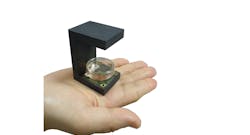
GRENOBLE, FRANCE. Miniaturization of sophisticated technologies has opened the door to innovative health-and-safety equipment that enables advances such as onsite, immediate evaluation of environmental risks and patient-sample analysis outside the lab. French research institute CEA-Leti’s lens-free microscope for diagnosing spinal meningitis at a fraction of the cost of bulky existing systems is the latest example in this field.
The microscope, which will be demonstrated at CES 2020 Jan. 7-10 in Las Vegas, can be operated by healthcare professionals at the patient’s bedside. It provides immediate results and accurate countings of white blood cells (leukocytes) in cerebrospinal fluid, which is required to diagnose spinal meningitis, an acute inflammation of the membranes covering the brain and spinal cord.
The technology costs about one-tenth as much as an optical microscope and can image up to 10,000 microscopic biological objects at a time. Protected by 25 patents, the microscope has neither lenses nor moving parts. The analysis results are available in about one minute.
The system operates with a near-infrared light emitted by a LED that is diffracted by the biological sample being analyzed to generate a holographic pattern captured by a CMOS image sensor. Holographic reconstruction algorithms digitally recreate the image of the object on a display. Artificial-intelligence software then detects, analyzes, and even classifies biological objects by tracking metrics of interest. All of these steps are automated.
CES 2020 demo
Visitors at CEA-Leti’s booth at Eureka Park-Sands Expo can observe the power of lens-free imaging to count and identify a variety of biological objects. Moving the slide under the microscope, they will see the image being digitally reconstructed in real time by the microscope’s algorithms. And they can watch as the embedded AI algorithms count and classify the objects being observed (eukaryotic cells, colon or lung tissues, plankton, neurons, etc.) in real time.
Find more details here.
